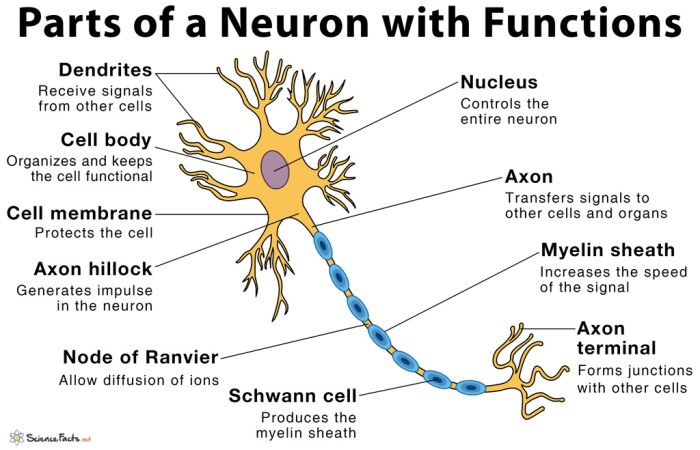
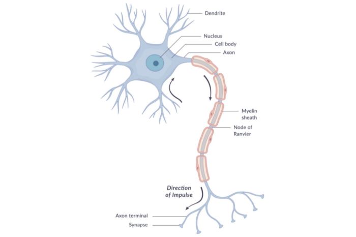
Figure 7-1 is a diagram of a neuron – Figure 7-1, a diagram of a neuron, serves as a foundational representation of the intricate structure and function of these fundamental units of the nervous system. This diagram provides a comprehensive overview of the neuron’s morphology, electrical properties, and role in chemical communication, making it an invaluable tool for understanding the fundamental principles of neuroscience.
Delving into the diagram, we embark on an exploration of the neuron’s structural components, including the cell body (soma), dendrites, and axon. We will elucidate the function of each component, unraveling the intricate mechanisms that govern neuronal communication and information processing.
Introduction
Figure 7-1 presents a comprehensive diagram of a neuron, the fundamental building block of the nervous system. Understanding the structure and function of neurons is crucial for comprehending neural communication and the workings of the brain.
Neurons are specialized cells that transmit electrical and chemical signals to communicate information throughout the nervous system. They consist of a cell body, dendrites, and an axon, each with distinct roles in signal processing and transmission.
Structure of a Neuron
Cell Body (Soma)
The cell body, or soma, is the metabolic center of the neuron. It contains the nucleus, which houses the genetic material, and other organelles responsible for protein synthesis and cellular maintenance.
Dendrites
Dendrites are short, branched extensions of the cell body that receive signals from other neurons. They are covered in specialized receptors that bind to neurotransmitters, the chemical messengers that facilitate communication between neurons.
Axon
The axon is a long, slender projection that extends from the cell body and transmits signals away from the neuron. It consists of the axon hillock, the initial segment where action potentials are generated, the myelin sheath, which insulates the axon and speeds up signal propagation, and the terminal buttons, where neurotransmitters are released.
Electrical Properties of a Neuron
Resting Membrane Potential
When a neuron is at rest, it maintains a negative electrical potential across its membrane, known as the resting membrane potential. This potential is maintained by the unequal distribution of ions across the membrane, with more sodium ions outside the cell and more potassium ions inside.
Action Potential Generation, Figure 7-1 is a diagram of a neuron
When a neuron receives a strong enough stimulus, it triggers an action potential, a brief electrical impulse that travels along the axon. Action potentials are generated when voltage-gated ion channels in the axon hillock open, allowing sodium ions to rush into the cell, depolarizing the membrane.
This depolarization triggers the opening of more ion channels, creating a self-propagating wave of depolarization that travels down the axon.
Propagation of Action Potentials
Action potentials are propagated along the axon due to the influx of sodium ions and the subsequent outflow of potassium ions. The myelin sheath surrounding the axon insulates it, preventing ion leakage and allowing for faster propagation.
Chemical Communication between Neurons: Figure 7-1 Is A Diagram Of A Neuron
Neurotransmitters
Neurotransmitters are chemical messengers that facilitate communication between neurons. They are released from the terminal buttons of the presynaptic neuron and bind to receptors on the dendrites of the postsynaptic neuron, triggering a response.
Neurotransmitter Release and Reuptake
Neurotransmitters are released from the presynaptic neuron by a process called exocytosis. Once released, they diffuse across the synaptic cleft and bind to receptors on the postsynaptic neuron. After binding, neurotransmitters are either reabsorbed by the presynaptic neuron or broken down by enzymes.
Types of Neurotransmitters
There are numerous neurotransmitters, each with its own specific function. Some common neurotransmitters include glutamate, GABA, dopamine, serotonin, and norepinephrine.
Plasticity and Learning

Synaptic Plasticity
Synaptic plasticity refers to the ability of synapses, the junctions between neurons, to strengthen or weaken over time. This plasticity is the basis for learning and memory.
Types of Synaptic Plasticity
There are two main types of synaptic plasticity: long-term potentiation (LTP) and long-term depression (LTD). LTP occurs when a synapse is repeatedly activated, leading to an increase in its strength. LTD occurs when a synapse is infrequently activated, leading to a decrease in its strength.
Learning and Memory
Synaptic plasticity plays a crucial role in learning and memory. LTP is associated with the formation of new memories, while LTD is associated with the forgetting of old memories.
Applications in Neuroscience

Neurological Disorders and Diseases
Understanding the structure and function of neurons is essential for comprehending neurological disorders and diseases. Figure 7-1 can help identify and localize malfunctions in neurons, aiding in the diagnosis and treatment of conditions such as Alzheimer’s disease, Parkinson’s disease, and epilepsy.
Development of Treatments
The knowledge gained from studying neuron structure and function has led to the development of treatments for neurological conditions. For example, drugs that target ion channels can be used to treat epilepsy, and drugs that enhance synaptic plasticity can be used to treat memory disorders.
Advances in Neuroscience
The study of neuron structure and function has revolutionized our understanding of the brain and its workings. This research has provided insights into the mechanisms of learning and memory, the basis of consciousness, and the development of novel treatments for neurological disorders.
FAQ Section
What is the significance of figure 7-1?
Figure 7-1 is a diagram that provides a comprehensive overview of the structure and function of a neuron. It serves as a foundational representation of the fundamental unit of the nervous system.
What are the key structural components of a neuron?
The key structural components of a neuron include the cell body (soma), dendrites, and axon. Each component plays a specific role in the neuron’s function.
How do neurons communicate with each other?
Neurons communicate with each other through chemical communication. Neurotransmitters are released from the presynaptic neuron and bind to receptors on the postsynaptic neuron, triggering a response.
What is synaptic plasticity?
Synaptic plasticity is the ability of synapses to change their strength over time. This process is essential for learning and memory.